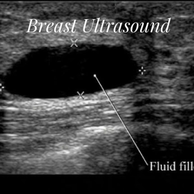
Institute of Ultrasound training is offering online and offline courses in Beast ultrasound in India. We have best expert of ultrasound training and providing certificate and diploma courses in Breast Ultrasonography. We use latest technology of radiology in India. We are offering diploma of various university as well as International university. We have online classes of breast ultrasound training. We are also offering clinical expertise for our medical students in India.
What is Breast Ultrasound?
Ultrasound imaging of the breast creates images of the inside anatomy of the breast by using sound waves. It is generally used to aid in the diagnosis of breast masses or other abnormalities discovered by a physical exam, mammography, or breast MRI. Ultrasound is non-invasive, safe, and does not use radiation.
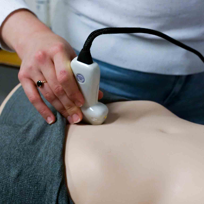
This method takes little to no preparation. Wear loose, comfy clothes and leave your jewels at home. During the process, you will be requested to undress from the waist up and don a robe. Ultrasound is non-invasive and painless. It uses sound waves to create images of the interior of the body. Ultrasound imaging is also known as sonography or ultrasound scanning. It employs a tiny probe known as a transducer and gel that is applied directly to the skin. High-frequency sound waves flow from the probe into the body via the gel. The noises that bounce back are collected by the probe. These sound waves are used by a computer to generate a picture. Ultrasound examinations do not involve the use of radiation (as used in x-rays). Images recorded in real-time can reveal the structure and movement of the body's interior organs. They can also demonstrate blood flow through blood vessels. Ultrasound imaging is a non-invasive medical diagnostic that aids doctors in the diagnosis and treatment of medical disorders.Doppler ultrasonography is a kind of ultrasound that measures the movement of materials in the body. It lets the clinician to examine and analyse blood flow via the body's arteries and veins.Breast ultrasound imaging creates an image of the interior tissues of the breast.
The sonographer or physician doing the test may employ Doppler methods to measure blood flow or lack of flow in any breast mass during a breast ultrasound examination. This may offer more information on the cause of the mass in some circumstances.Identifying the Type of Breast Abnormality.Breast ultrasonography is primarily used to aid in the diagnosis of breast abnormalities discovered by a physical exam (such as a lump) and to define probable abnormalities observed on mammography or breast magnetic resonance imaging (MRI).Ultrasound imaging can assist detect if an abnormality is solid (a non-cancerous mass of tissue or a malignant tumour), fluid-filled (a benign cyst), or both cystic and solid.
Doppler ultrasonography is used to evaluate blood supply in breast tumours.Breast Cancer Screening Supplement
Mammography is the only breast cancer screening method that has been shown to minimise breast cancer fatalities by early diagnosis. Nonetheless, mammography do not identify all types of breast cancer. On mammography, certain breast lesions and abnormalities are not evident or are difficult to interpret. Breasts that are considered dense have a lot of glandular and connective tissues and not much fatty tissue, and that makes cancer harder to detect. Many studies have demonstrated that ultrasound and magnetic resonance imaging (MRI) can assist complement mammography by identifying breast tumours that mammography may miss. Your doctor can advise you on whether one of these tests is right for you. Although MRI is more sensitive than ultrasound in detecting breast cancer, it is not available to all women. Screening MRI is not required if screening MRI is conducted; nevertheless, ultrasonography may be utilised to define and biopsy anomalies observed on MRI. When ultrasonography is used for screening, abnormalities that are not evident on mammography can be found, including those that may necessitate biopsy. Many of the anomalies discovered A physician may opt to do an ultrasound-guided biopsy if an ultrasound examination detects a suspected breast anomaly. Ultrasound is frequently used to guide biopsy operations since it gives real-time visuals. An ultrasound evaluation is typically required prior to the biopsy in order to plan the surgery and evaluate whether this kind of biopsy may be employed. It is best platform query related to Institute of Breast Ultrasound Courses is providing program of Breast Ultrasound Course for Surgeons, Breast Imaging Fellowship, Breast Sonography Course, Breast Ultrasound Training, Breast Ultrasound Certification for Mammographers, Ultrasound Breast Bioscopy Course, Breast Ultrasound Technologist, Breast Ultrasound Radiology Course and Diploma in Breast Ultrasound in India.
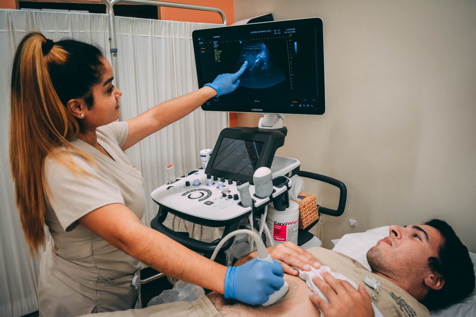 Diploma in Ultrasonography
 Infertility Ultrasound Course
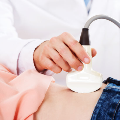 Abdominal Ultrasonography Course
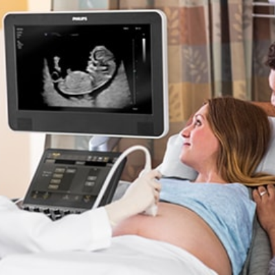 Obstetrics & Gynaecology Ultrasound Training Course
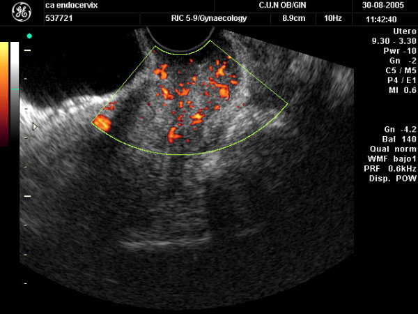 Certificate Course in Level II Scan + Color Doppler in Obstetric & Gynaecology
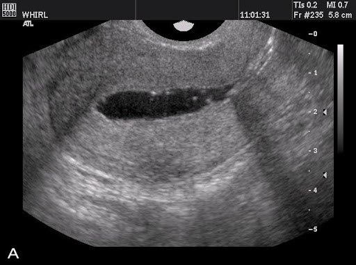 Certificate Course in Infertility Ultrasound + TVS
 Sonography Course
 Sonology Course
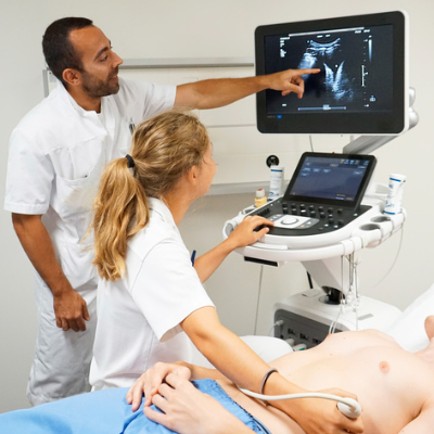 Ultrasonography Course
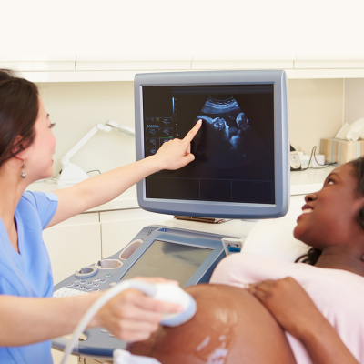 USG Course
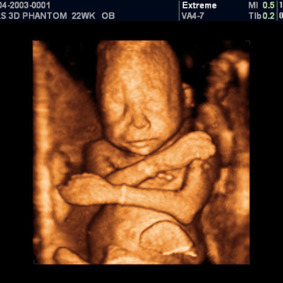 Fetal Ultrasound Training Course
 Ultrasound Training Course
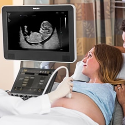 Gynaecology Ultrasound Courses
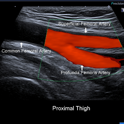 Vascular Ultrasound Courses
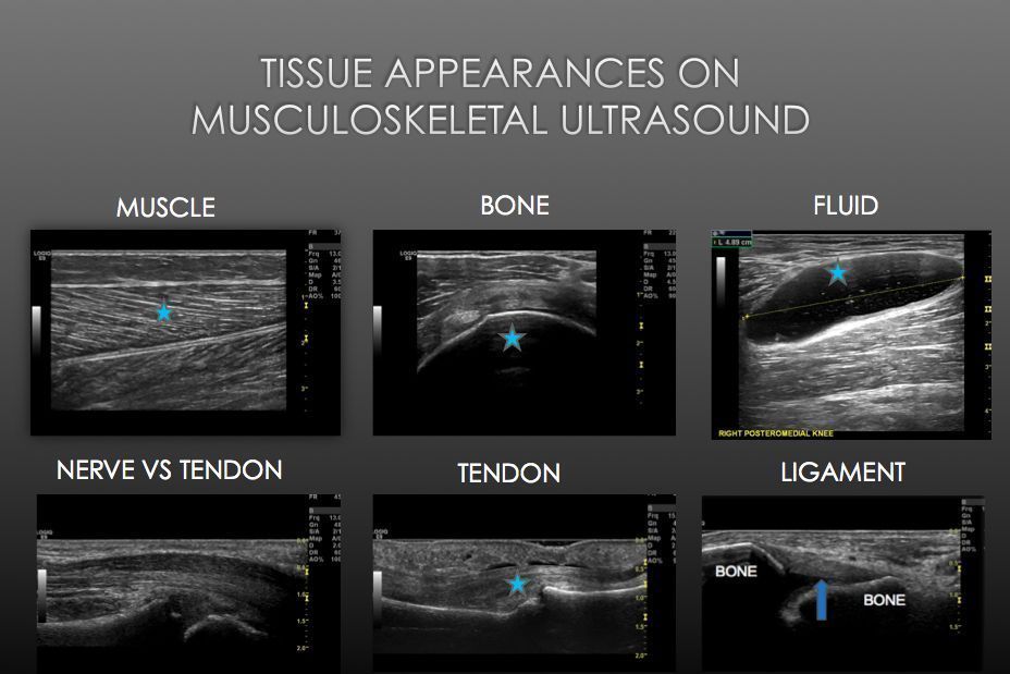 Musculoskeletal Ultrasound Course
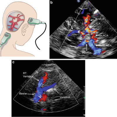 Transcranial Doppler Ultrasound
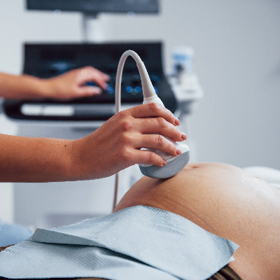 Obstetric Ultrasound Courses
 OBS and Gynae Ultrasound Course
 Certificate in Gynecology and Obstetrics
 2D Echo Ultrasound
 DGO Course